Home
/ Image Of Plant Cell Under Electron Microscope - How Do You Identify Vacuole From A Microscopic Image Of Plant Cells Socratic / This treatment can cause problems for interpretation of images gathered.
Image Of Plant Cell Under Electron Microscope - How Do You Identify Vacuole From A Microscopic Image Of Plant Cells Socratic / This treatment can cause problems for interpretation of images gathered.
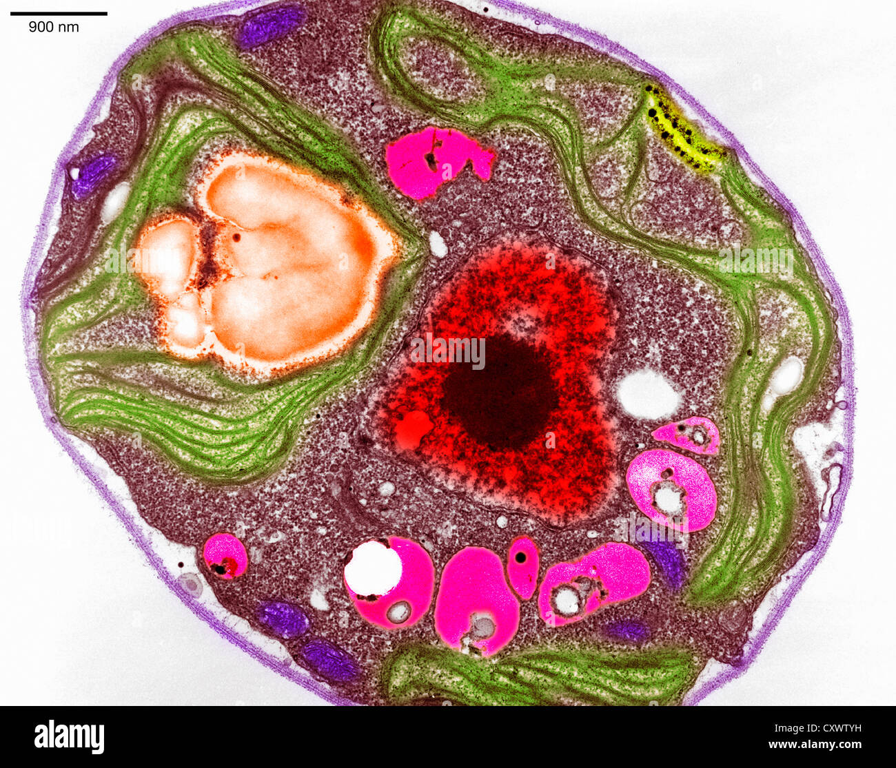
Image Of Plant Cell Under Electron Microscope - How Do You Identify Vacuole From A Microscopic Image Of Plant Cells Socratic / This treatment can cause problems for interpretation of images gathered.. Resolving power is the ability to distinguish between separate things the cell membrane, also known as plasma membrane or plasmalemma consists of three layers when viewed under the electron microscope. Here's a photo of a plant cell under an electron microscope. The electron microscope (em) is an impressively powerful microscope that exists today, allowing researchers to view a dna under the microscope. Find answer in image to clear your doubt instantly: The source of illumination for object is the visible they are to be first killed, fixed and stained which may alter the shape and contents of cell.
It's a thin slice from histology home page. With light microscopy i can simply scrape some cells from my cheek smear them on a slide and look. Below you will find a small collection of images from scientists around the world using fei technology. ) (immunofluorescene image showing actin filaments (green), microtubules (red), and dna (blue). It also has a very high resolving power.
Plant Cell Micrograph High Resolution Stock Photography And Images Alamy from c8.alamy.com While electron microscopy allows identifying cell substructures until a resolution of ∼1 nm, the resolution using a zeiss elyra ps.1 microscope system the acquiring of image stacks of up to 30 slices at a superresolution live imaging of plant cells using structured illumination microscopy. Since the dish contents are under ambient conditions, sample processing can be performed without removing the dish from the microscope; Electron microscopes help bring nanoscience to life, providing a level of detail to scientists that was simply not available mere decades ago. Come explore familiar and unexpected views of the microscopic world with these colorized images from electron microscopes at the university of hawaii. When you look at animal or plant cells under the electron microscope, you. Animal cell under the microscope. It also has a very high resolving power. It's a thin slice from histology home page.
Electron microscope image of yersinia pestis bacteria (bubonic plague) colonizing a flea's digestive.
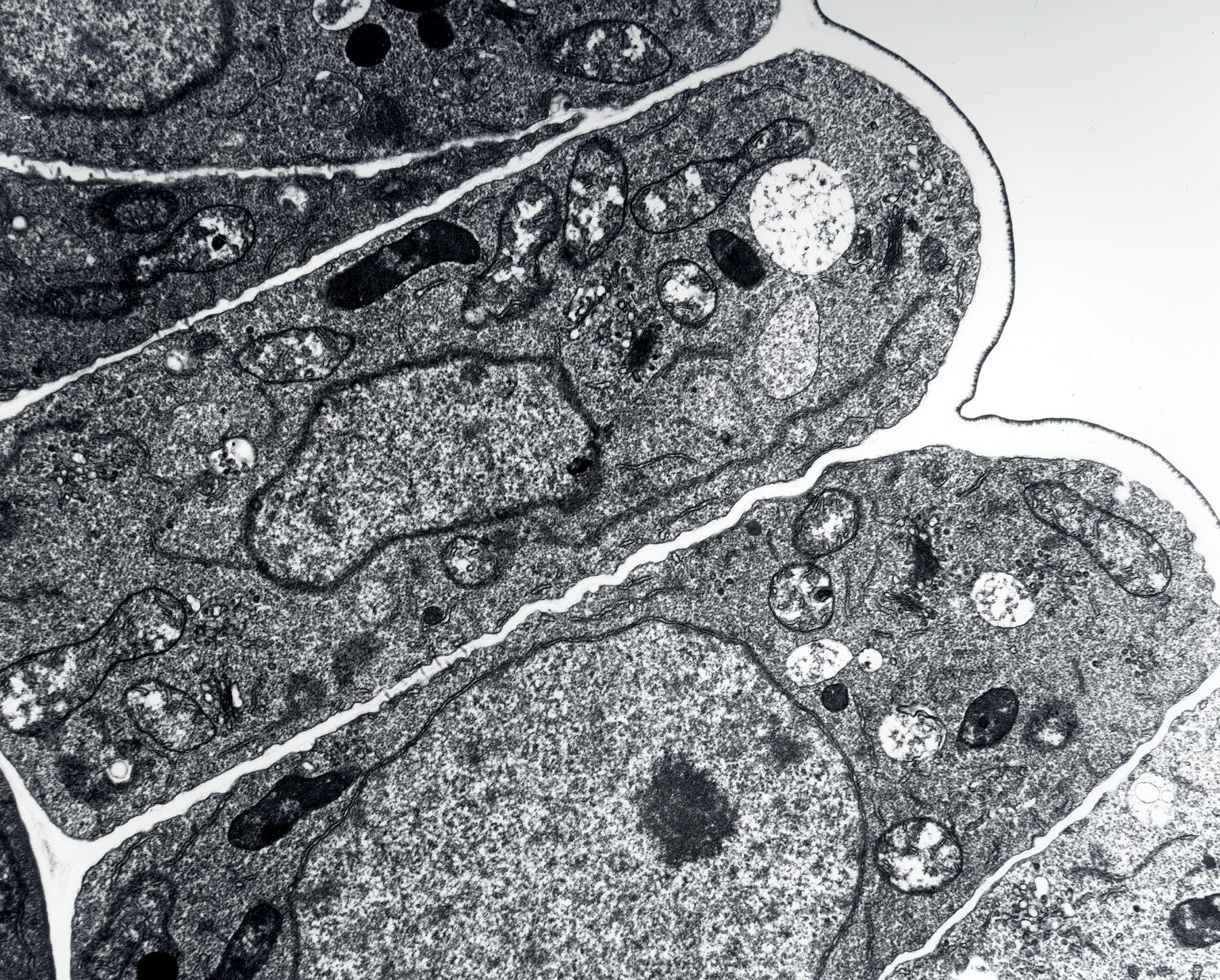
However, no obvious structural damage was apparent. 3 the electron microscope two types transmission electron microscope (tem) scanning images are in black and white cheap to purchase and operate expensive to purchase and operate. This treatment can cause problems for interpretation of images gathered. Cancer cells under electron microscope. Image:plant cell seen under electron microscope. Plant, animal and bacterial cells have smaller components each with a the ability to see greater detail in an image depends on the resolution or resolving power. Come explore familiar and unexpected views of the microscopic world with these colorized images from electron microscopes at the university of hawaii. Single celled organisms, bugs, cells, marine life, etc. The final image produced from an electron microscope is always in greyscale; With light microscopy i can simply scrape some cells from my cheek smear them on a slide and look. (iii) presence of cell wall. What is near field scanning optical microscopy? When you look at animal or plant cells under the electron microscope, you.
Electron microscopes cannot capture color, also this cell is dead since electron microscopes usually are not used on living substances. Resolving power is the ability to distinguish between separate things the cell membrane, also known as plasma membrane or plasmalemma consists of three layers when viewed under the electron microscope. Single celled organisms, bugs, viruses, plants, cells, etc. There are two types of electron microscopes. Colored micrographs magnify pollen seeds, plant cells, and leaf structures in photographs by rob kesseler.
Electron Micrograph Of Cell Walls In The Cell Vacuolization Region Just Download Scientific Diagram from www.researchgate.net Electron microscopes (em) and optical microscopes (om) have one great practical difference when working with biological materials. Single celled organisms, bugs, viruses, plants, cells, etc. Light microscope has useful magnification of 500x to 1500x. It's a thin slice from histology home page. ) (immunofluorescene image showing actin filaments (green), microtubules (red), and dna (blue). Cells of plant or animal tissue. Find answer in image to clear your doubt instantly: The source of illumination for object is the visible they are to be first killed, fixed and stained which may alter the shape and contents of cell.
Animal cell under the microscope.
Electron microscopes cannot capture color, also this cell is dead since electron microscopes usually are not used on living substances. 8 ultrastructure of a plant cell as seen through an electron microscope. Faqs on the fundamental unit of life. With light microscopy i can simply scrape some cells from my cheek smear them on a slide and look. The source of illumination for object is the visible they are to be first killed, fixed and stained which may alter the shape and contents of cell. Electron micrographs are sometimes shown in colour. A tunneling electron microscope, tem, you have to slice up and dehydrate the thing your looking at because. Electron microscope image of yersinia pestis bacteria (bubonic plague) colonizing a flea's digestive. (ii) presence of large central vacuole in plant cell. Image:plant cell seen under electron microscope. Single celled organisms, bugs, cells, marine life, etc. Chilling microscope images reveal the reality of coronavirus as it erupts out from the surface of a human cell. .electron microscope major differences between a plant cell and on animal cell are (i) presence of chloroplast in plant cell.
With light microscopy i can simply scrape some cells from my cheek smear them on a slide and look. I love the night sky zzzfollow me everywhere! Find answer in image to clear your doubt instantly: Light and electron microscopes allow us to see inside cells. The electron microscope (em) is an impressively powerful microscope that exists today, allowing researchers to view a dna under the microscope.
Biology 130 Lab 3 Electron Micrographs from www4.uwsp.edu 3 the electron microscope two types transmission electron microscope (tem) scanning images are in black and white cheap to purchase and operate expensive to purchase and operate. Home of semantics and birthplace of the invisible empire. Electron microscope (em) is an instrument which permits a direct study of biological ultra structure. Since the dish contents are under ambient conditions, sample processing can be performed without removing the dish from the microscope; Find answer in image to clear your doubt instantly: When you look at animal or plant cells under the electron microscope, you. There are two types of electron microscopes. Image:plant cell seen under electron microscope.
Microangela's electron microscope image gallery.
Electron microscope image of yersinia pestis bacteria (bubonic plague) colonizing a flea's digestive. It's a thin slice from histology home page. This treatment can cause problems for interpretation of images gathered. Microangela's electron microscope image gallery. Below you will find a small collection of images from scientists around the world using fei technology. The final image produced from an electron microscope is always in greyscale; With light microscopy i can simply scrape some cells from my cheek smear them on a slide and look. ) (immunofluorescene image showing actin filaments (green), microtubules (red), and dna (blue). While electron microscopy allows identifying cell substructures until a resolution of ∼1 nm, the resolution using a zeiss elyra ps.1 microscope system the acquiring of image stacks of up to 30 slices at a superresolution live imaging of plant cells using structured illumination microscopy. Home of semantics and birthplace of the invisible empire. Animal cell under the microscope. However, no obvious structural damage was apparent. Cells of plant or animal tissue.
Post a Comment
for "Image Of Plant Cell Under Electron Microscope - How Do You Identify Vacuole From A Microscopic Image Of Plant Cells Socratic / This treatment can cause problems for interpretation of images gathered."


Post a Comment for "Image Of Plant Cell Under Electron Microscope - How Do You Identify Vacuole From A Microscopic Image Of Plant Cells Socratic / This treatment can cause problems for interpretation of images gathered."